
A Review On Cad Cam In Dentistry
INTRODUCTION
Over the past 30 years, CAD CAM (computer aided design computer aided machine) has gained popularity and confidence of the profession and patients alike. It has provided ease, comfort, and quality of restoration to both the dentists and dental lab technician. Moreover, restorations which are being produced through cad cam these days, are more durable, more marginally adaptive, more esthetically pleasing and more faster in fabrication as compared to the conventional restorations. But despite of these advantages, cost is the major issue which has limited its use. Because of the financial constraints, in the under developed countries, dentists are still reluctant in using cad cam However, one should not overlook the benefits which CAD CAM provides. Accuracy and quality of CAD CAM in producing inlays, on lays, implant abutments, fixed partial dentures, crowns and bridges is acceptable and improving every day. This review article highlights the history and current indications of CAD CAM. It also provides information that how it works and what can be itsadvantages and disadvantages.
HISTORY OF CAD CAM
The history of dentistry is as old as human civilization in the ancient times. Hippocrates and Aristotle, have written many things about dental diseases and their treatments.1 In the eastern world, ancient Chinese literature is available regarding silver pastes (primitive amalgam). As the civilization was getting developed, newer and newer findings kept on becoming the part of dentistry. Commercial production of porcelain teeth by Samuel Stockton, introduction of amalgam by Crawcours and origination of cohesive gold foil method by Robert Arthur all have brought revolution in the field of dentistry.1 So revolutionization has become the habit of dentistry.
The history of cad cam itself is old. In ancient Egypt, Greece, and Rome, evidences suggest that cad cam was being practiced there even. Even Leonardo di Vinci has shown in his works, the use of modern graphics convention.2 But Euclid of Alexandria, the great mathematician, can truly be regarded as the person behind the modern CAD soft ware because today’s CAD software is based on Euclid’s axioms and postulates, which laid the foundation of Euclidian geometry.1,4 Iven Sutherland in early 1960s, developed the CAD software (sketchpad) but before that Dr. Patrick J. Hanratty, had already designed first numerically controlled CAM that was named as Pronto. So Dr Patrick is called as Father of CAD CAM.3,4
The introduction of CAD CAM in dentistry begins with the work of Dr Duret. He took optical impression of an abutment tooth after cutting and then with the help of that impression he fabricated a crown by using numerically control machine. He did that miracle in the field of dentistry in the year 1971.5 Later on Dr Duret along with his team mates, worked on Sopha system and introduced it but due to few reasons that system was not generally accepted by the people.6 Mormann was the person who introduced first commercially designed CAD CAM system in the year 1985 and given the name of CEREC to that system.7 He fabricated an inlay from a ceramic block after taking digital impression from intra oral camera. That was bit easy as compared to take impression of a prepared abutment tooth. Now this system is being used all over the world successfully for the fabrication of crowns, inlays, onlays etc. Dr Andersson developed Procera system in the mid-1980s and worked on titanium as a substitute of nickel chromium alloys.8 Previously, gold alloys were being used, but due to increase in prices of gold, people had to think of substitutes. Dr Andersson came up with titanium because allergies were reported with nickel chromium.
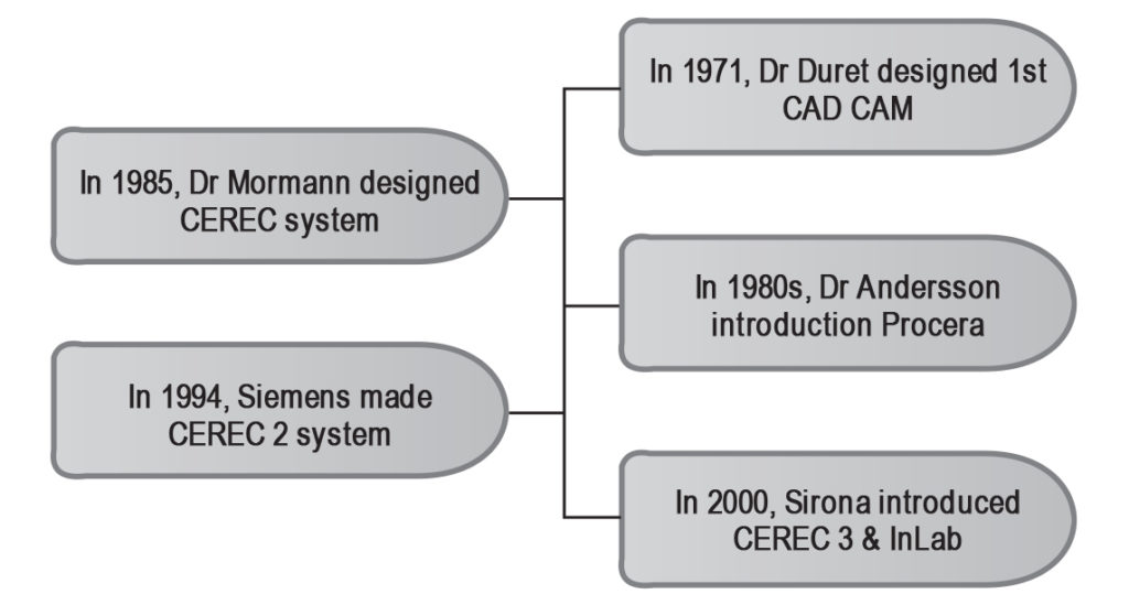 Fig 1: Summarizes the major advancements in CAD CAM.
CEREC 2 was introduced in 1994 by Siemens. This system was based on two dimensional principles and capable of producing inlay, onlay, veneers, partial & full crowns and copings. Currently 3rd generation of CEREC is in use, which is capable of producing inlay, onlay, veneers, partial & full crowns, copings as well as virtual automatic occlusal adjustment. This system was introduced by Sirona in 2005. This system is basically the advanced form of CEREC 3 which was earlier introduced in 2000 by Sirona but that system worked on two dimensional principles and was not able to provide virtual automatic occlusal adjustments.9
Types of CAD CAM Production
CAD CAM restorations can be produced by three different ways.
- Chair side production
- Laboratory production
- Centralized production
- Chair side production:
It involves taking an impression chair side and then producing the restoration at the same time. It doesn’t require involvement of the laboratory and the patient/s can have their restoration at the same appointment10. Obviously it saves times but it is expensive and puts extra cost on the patient.
2. Laboratory production
It is somewhat similar to the conventional method. The dentist takes the impression & sends it to the lab, where other works carried out by the lab technician.
3. Centralized production
In centralized production of the restoration, an impression is taken and a master cast is digitized in the lab, then it sends to outsource laboratory through internet. In that outsource lab, the final restoration is fabricated which sends back to the dentist. The idea is right in the sense that it requires digitizer and software only to perform the initial steps and a high quality restoration.10
METHODS OF DATA ACQUISITION IN CAD CAM
There are several methods by which data can be collected for CAD.
1) Intra oral scanning
2) Contact and non – contact digitization
3) Ct scan or MRI
Fig 2

1. Intra Oral Scanning
There are several intra oral scanners available in market namely CEREC® – by Sirona Dental System GMBH (DE), Lava C.O.S. – by 3M ESPE (US), iTero – by CADENT LTD (IL) and E4D – by D4D
TECHNOLOGIES, LLC (US) etc. What these systems do, they take 3-D virtual picture of the prepared tooth/teeth and the adjacent structures, directly into the patient’s mouth Later on these images transfer to the CAD for designing of the prosthesis.11 A comparison of intra oral scanning which is also called as digital impression and conventional impression is given below.
But despite of the advantages of digital impression technique, there are certain things which need to be considered while taking digital impressions. The dentist, who is taking intra oral pictures, must take care of moisture control. Soft tissue retraction and hemostasis should be considered too.12
2. Contact & Non – Contact Digitizing
This method involves taking an conventional impression and then after producing a model, transferring of that data into CAD through probe digitization ( contact) or laser light ( non – Contact ). In contact digitizing or scanning, a contact probe reads the anatomy of the model by following the contour of the physical structure. In non-contact scanning, apart from laser light, optics and charged -coupled devices are also used. Obviously, this technique doesn’t require any physical contact with the model, but precision in recording the details is required. It is assumed that, in comparison to contact scanning, this technique is relatively quick in gathering the data.13
Ct scan /MRI:
Computed Tomography (CT & Magnetic Resonance Imaging (MRI) are newer techniques for data acquisition for CAD Cam.By this method, individual images can be taken and then can be transferred to CAD. CT scan utilizes radiation for its data acquisition but MRI doesn’t. For soft tissue modelling, MRI data is suitable and for hard tissue (bone) modelling, CT data can be used.14
USES OF CAD CAM IN DENTISTRY
CAD CAM is being used in almost every field of dentistry these days. Following are its some of the uses in different specialties of dentistry:
Prosthodontics:
Whether it is removable or fixed prosthodontics, fabricating them through CAD CAM is becoming the choice of almost every leading dentist.
a) Removable Complete Dentures:
A group of Japanese investigators named Maeda et al, published a report in 1994, in which they proposed that removable complete dentures can be fabricated using CAD Cam technology.15 Since then many researches have been proposed regarding fabrication of removable complete dentures through CAD CAM but no clinical reports or trials have been published yet. Only 2 of the manufacturers claim that they manufacture RCD through CAD CAM.16
b) Removable Partial Dentures
Partial denture framework can be produced through CAD CAM by using additive prototyping technique.17,18
c) Crowns / Bridges
Zirconia is the widely used material for the fabrication of crown and bridge through CAD CAM. However, metal & porcelain crown & bridge can also be fabricated through CAD CAM. A study suggests that zirconia exhibits acceptable clinical results.19
d) Inlay, Onlay & Veneers
These restorations are also being produced through CAD CAM.20 A study says that inlays & onlays, produced through CAD CAM, have a higher survival rate.21
Orthodontics
In orthodontics, clear aligners have gained so much popularity due to esthetics. Now patients don’t have to show metallic brackets and wires. The credit goes to CAD CAM again.22 Also lingual bracket system is in demand due to its invisibility. The wires and bracket system for the lingual bracket system is fabricated through CAD CAM.23 Orthodontic mini implants can also be positioned using CAD CAM technology.24
Implant Dentistry:
Implant abutments and surgical guides for the placement of implants are being produced through CAD CAM these days.25
Maxillo-Facial Prosthetics:
A study shows the difference in between the outcome of the artificial ear prosthesis carved by hand & fabricated through CAD CAM. The latter is superior over the hand carved prosthesis.26 So artificial ear27, artificial nasal prosthesis28 are being produced through CAD CAM. Not only this, but certain bony defects following trauma or removal of tumors are also being treated by manufacturing implants through CAD CAM.29
ADVANTAGES AND DISADVANTAGES OF CAD CAM
If we compare the advantages of CAD CAM restorations over the conventional one, we will definitely place CAD CAM restorations on top. They provide us quality restorations with quick and easy fabrication. Scanning of intra oral tissues takes less time than conventional impression, and if the chair side system is available, the patients can get their restorations in one appointment.Quality of these restorations has been demonstrated in so many studies.
But despite of these advantages, cost is still a major issue. Also taking digital impression is a big challenge for the dentists, because they have to take care of soft tissue retraction, moisture control etc.
CONCLUSION
CAD CAM has no doubt changed the concept of modern world dentistry. Different specialties of dentistry are being successfully benefited by CAD CAM. Either orthodontics or prosthodontics, quality treatment is possible with accuracy and effectiveness. But cost of these treatments is still a problem for the patients and the dentists, esp. of the developing countries. We are hopeful that in the near future, CAD CAM will be started using widely in the developing countries as well.
Author Contribution: UBI conceived the idea and made the initial draft, KA revised the manuscript and RN collected the references. Disclosure: None disclosed
REFERENCES
- ada.org/en/about-the-ada/ada-history-andpresidents-of-the-ada/ada-history-of-dentistry-timeline. Accessed 10-10-2015.
- Zeid, Ibrahim,Cad/Cam Theory and Practice,1991, McGraw-Hill.
- http://www.cadazz.com/cad-software-history.htm
- ukessays.com.
- Duret F, Preston JD. CAD CAM imaging in dentistry. Curr Opin Dent 1991;1(2):150-154.
- T Miyazaki, Y Hotta.CAD/CAM system available for the fabrication of crown and bridge restorations.Aust Dent Jour. 2011;56:97-106.
- Mormann WH, Brandestini M, Lutz F, Barbakow F.Chairside Computer Aided Direct Ceramic Inlays. Quintessence Int 1989;20:329-339.
- T Miyazaki, Y Hotta, J Kunii, S Kuriyama, Y Tamaki.A review of dental CAD CAM: Current status and future perspectives from 20 years of experience.Dent Mat Jour 2009;28:44-56.
- WH, Mormann. The evolution of the CEREC system.J Am Dent Assoc 2006;137:7S-13S.
- Beuer, J. Schweiger, D. Edelhoff.Digital Dentistry: an overview of recent developments for CAD/CAM generated restorations. Br Dent J 2008;201:505-511.
- A Kilpeta, M Capni, L Governu, L Blois.A comparative analysis of intra oral 3D digital scanner for restorative dentistry. The Int Jour Of Med Tech 2008;5.
- Gary Davidowitz, Philip G. Kotick. The Use Of CAD /CAM in dentistry. Dent Clin N Am 2011;55:559570.
- V Raja, KJ Fernandes.Reverse Engineering: An Industrial Perspective 2008. Springer.
- L Siva Rama Krishna, Manmadhachary, P. Bal Reddy, Sriram Venkatesh.Development of optimum preplanning for maxillofacial surgery using selective laser sintering.International J Eng Sci Tech 2011;3:185-196.
- Maeda Y, Minoura M, Tsutsumi S, Okada M, Nokubi T. A CAD CAM system for removable denture.Part 1: Fabrication of Complete denture.Int J Prosthdont 1994;7:17-21.
- Avinash S. Bidra, Thomas D. Taylor, John R. Agar.Computer -Aided Technology For Fabricating Complete Dentures: Systematic Review Of Historical Background, Current Status And Future Perspectives.JProsth Dent 2013;109:361-366.
- Chatham C, Spencer MH, Wood DJ, Johnson A. The introduction of digital dental technology into BDS curricula. Br Dent J. 2014:5;217:639-42.
- Williams RJ, Bibb R, Eggbeer D, Collis J. Use of CAD/CAM technology to fabricate a removable partial denture framework. J Prosthet Dent. 2006;96:96-99.
- Batson ER, Cooper LF, Duqum I, Mendona G. Clinical outcomes of three different crown systems with CAD/CAM technology. J Prosthet Dent. 2014;112:770777.
- TrushkowskyRD.Ceramic inlay fabrication with three dimensional copy milling technology Celay.Compend Cont Edu Dent 1998;19:1077-80.
- Otto T, De Nisco S.Computer aided direct ceramic restorations: A 10 year prospective clinical study of CEREC CAD/CAM inlays and onlays. Int J Prosthodont 2002;15:122-128.
- Aristides BM. Advances in digital technology and orthodontics: A reference to the invisalign method.Med Sci Monit 2005;11:139-142.
- Wiechmann D, Rummel V, Thalheim A, Simon JS, Wiechmann L.Customized Brackets And Arch wires For Lingual Orthodontic Treatment.Am J Orthod Dentofac Orthop 2003;124:593-599.
- Liu H, Liu D, Wang G, Wang C, Zhao Z.Accuracy of surgical positioning of orthodontic mini screws with a computer-aided design and manufacturing template. Am JOrthod Dentofac Orthop 2010;137:728e1-28e10.
- . Fuster-Torres MA, Albalat-Estela S, Alca±iz-Raya M, Pe±arrocha-Diago MCAD CAM dental systems in implant dentistry:Update. Med Oral Patol Oral Cir Bucal 2009;14:E141-145.
- Sykes LM, Parrott AM, Owen CP, Snaddon DR. Applications of rapid prototyping in maxillofacial prosthetics. Int J Prosthodont 2004;17:454-459.
- Ciocca L, Scotti R.CAD CAM generated ear cast by means of laser scanner and rapid prototyping machine. J Prosthtic Dent 2204;92:591-5.
- Leonardo Ciocca, Massimilliano Fantini, Francesca De Crescenzio, Franco Persiani, Roberto Scotti.New protocol for construction of eye glasses- supported provisional nasal prosthesis using CAD CAM techniques. J Rehab Res Dev 2010;47:595-604.
- Eufinger H, Wehmoller M, Machtens E, Heuser L, Harder A, Kruse D.Reconstruction of craniofacial bone defects with individual alloplastic implants based on CAD/CAM manipulated CT data.J Maxillofac Surg 1995;23:175-181.