
Root Canal Treatment of Maxillary Premolar In A Subject with Retention Cyst in Maxillary Sinus: A Diagnostic Challenge
INTRODUCTION
The dental pulp is a neurovascular tissue that resides in a rigid chamber comprising of tooth enamel dentine and cementum. This rigid encasing not only provides mechanical support but also protects the pulp from biochemical insults and against microbial invasion as well. Therefore, it is important that the tooth structure maintains its structural integrity so that the dental pulp remains protected. Caries, cracks, fractures and open restoration margins provide pathways for microorganisms and their toxins to enter into the pulp.(1)
Toothache is one of the most common complaints seen in the dental clinics, and yet diagnosis of pulp disease is often difficult due to the seemingly unclear symptoms(1) Even in the presence of a detailed history and a thorough clinical examination, its diagnosis can be difficult, and can be further complicated by presence of pathology in surrounding anatomical structures and referred pain. A delay in diagnosis can lead to a delay in treatment therefore causing undue distress to the patient.
Non odontogenic pathology is sometimes difficult to diagnose and appears clinically as odoontogenic toothache, andvice versa. Especially if present in the vicinity of roots of teeth, and therefore the clinician may decide to defer treatment until localization of symptoms to be sure of odontogenic versus non odontogenic pain. Cracked teeth most often present with vague symptoms of pain and tenderness, reversible or irreversible pulpitic symptoms and are one of the more diffucltut entities to diagnose especially if present as partial or hidden cracks.
CASE REPORT
A 49 year old female presented to the Dental Clinics at Aga Khan University Hospital Karachi on August 15th 2011 with complaint of mild tenderness in the upper right premolar area on chewing since two weeks. There was no history of spontaneous pain or sensitivity to hot, cold or sweets. She also complained of heaviness in the right maxillary sinus area.
Her past medical history revealed that she had a history of chronic sinusitis, and took anti-allergic medication for allergies. She had a history of acute bronchitis in 2003, and was hospitalized in 2010 for dengue fever.
On her extra-oral examination, there were no facial asymmetries; mild tenderness on palpation in the right maxillary sinus area was noted. No palpable lymph nodes and no tenderness over the temporo-mandibular joint and muscle area were noted.
Intra oral examination revealed short clinical crowns, more square in shape. No carious lesion was seen. However all her third molars and left upper and both lower first molars were missing.The upper first molars # 26, 36 and 46 were extracted when she was 12 years old due to dental decay. She never got replacements done for these teeth. There was no tenderness on percussion in any of the teeth except teeth # 14, 15 and 16. Periodontal probing with BPE revealed a score of 1 in all sextants. There was no mobility seen in any tooth. Vitality testing of teeth # 14,15 1nd 16 was carried out with endo ice, warm gutta percha sticks and electric pulp testing. All methods revealed vitality status within physiological limits.
Radiographic examination revealed no abnormal findings; the lamina dura of the teeth in question was intact with adequate crestal bone levels. However, some calculus spurs were identified.
DIAGNOSIS
A definitive diagnosis could not be reached at that point in time, her occlusal contacts on teeth #13, 14 and 15 were adjusted, full mouth scaling and polishing was carried out and she was advised to come back for a follow up after 3 weeks time. She was also advised to consult with the ENT specialist for management of chronic sinusitis.
The patient consulted with ENT and was advised a Para-nasal Sinuses Radiograph (PNS view) and a cystic lesion in the right maxillary sinus area was identified. (See figure 2) She was prescribed antibiotics and nasal
Figure 2: PNS view advised by the ENT Specialist
decongestants and surgical drainage and resection if the symptoms did not subside.
The patient did not return for a follow-up until December 2011 with acute pain in her right maxillary first premolar area. She reported of worsening symptoms during the past four months with a decrease in her chewing efficiency on the affected side for which she was on multiple courses of antibiotics advised by her ENT and general physician. Periapical radiograph was repeated of tooth #13,14 and 15 and an apical radiolucency with tooth #14 was evident (Figure 3). The tooth was acutely tender to palpation, thermal testing
Figure 3: Peripaical radiograph 4 months after initial presentation
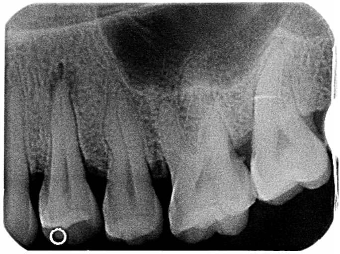
was repeated and the tooth was not responsive to any of the stimuli (cold, hot, EPT).
Mobility of this tooth was within normal limits (2mm) and periodontal probing depth was 3.5 mm.
The diagnosis made was Pulp necrosis secondary to crack tooth syndrome and the treatment plan advised was root canal treatment of tooth #14 followed by a crown, full mouth debridement and replacement of missing teeth by fixed or removable prostheses.
Root canal treatment was carried out and completed after which the symptoms resolved completely in 3 weeks time.
Figure 3: Immediate post operative image

3 months follow-up

DISCUSSION
Periapical lesions can be of endodontic or nonendodontic origin and may involve one or multiple adjacent teeth. Lesions of endodontic origin have been classified as periapical abscesses, granulomas, and cysts depending on their histological typing (2). Periapical lesions of non-endodontic origin can develop around the teeths periapical region and if not diagnosed early and correctly can expand to large lesions. Reports about nonendodontic lesions mimicking apical periodontitis and their misdiagnosis can be found frequently in the literature. (2)
Diagnosis is confirmed with the help of a detailed history and examination, periapical radiographs and electric and thermal pulp testing. However, two dimensional periapical radiographs often present with limitations in assessment of three dimensional root structures. Cone beam computed tomography is a better aid to reach a diagnosis in complicated cases and particularly could have added sufficient value in this case by disclosing any cracks or fractures and also in showing the link of the cystic lesion to the tooth. In this particular case, the first evaluation did not reveal any radiographic changes nor was the history or clinical examination guiding the clinicians toward a diagnosis In this particular case, the first evaluation did not reveal any radiographic changes nor was the history or clinical examination guiding the clinician towards a diagnosis. The patient had a history of chronic sinusitis, and therefore, was advised to seek an ENT evaluation as well. The patient did not return to clinic till 4 months after, when there was an abscess formed associated non vital # 14 which was extremely tender to palpation on presentation.
In this authors opinion, had the patient returned to clinic within 3-4 weeks of first evaluation, she would have been saved multiple visits to ENT, numerous courses of antibiotics and a visit to the emergency room as well. The question remains, whether it was lack of clinical judgment or lack of substantial evidence responsible for not formulating a definitive diagnosis earlier, or whether the endodontic pathology was below the diagnostic threshold on first presentation. In any case, the patient did not show up for 4 months after first examination till she got her upper premolar affected with abscess.
Infected root canal systems can lead to inflammation of periapical alveolar bone therefore leading to the development of apical periodontitis. When associated with maxillary canines and premolars, apical periodontitis can trigger inflammation of the mucosal lining of the maxillary sinus and lead to development of maxillary mucositis, periositits as well as sinusitis. Similarly, maxillary sinusitis can also present with symptoms of toothache and tenderness in the area of maxillary canines and premolars. This patient visited her
ENT frequently due to chronic sinusitis and kept getting treatment for such.
Other differentials that were suggestive of her symptoms were occlusal trauma leading to apical periodontitis, cracked tooth syndrome, and endoantral syndrome.
At her first visit, occlusal trauma was the most likely suspect due to multiple missing molars, mild pain only on chewing which lead to the dentist adjusted her heavy occlusal contacts on the affected side by means of superficial enameloplasty.
The normal mucosal lining within the maxillary sinus is <1 mm in thickness and not discernible radiographically. Sinus mucositis may develop in response to infectious or allergic stimuli producing a band of noncorticated mucosal tissue paralleling the bony wall of the sinus.(3-5) In some instances new bone formation is also seen when the noxious stimuli are odontogenic and travel through cancellous bone towards the corticated floor of the sinus.(3)
A diagnosis of endoantral syndrome could not be made at the second visit due to lack of communication and presence of corticated bone between the tooth and the floor of the right maxillary sinus.
The most likely diagnosis reached in the present case report was that of cracked tooth syndrome. Guersten et al. stated that excessive forces applied to a healthy tooth or physiologic forces applied to a weakened tooth can cause an incomplete fracture of enamel or dentine. (6) Cracks are not discernible on radiographs, even on multiple angulations of exposure. Cone beam computed tomography, and infrared thermography (7) are currently better modality to diagnose thin hairline fractures in teeth. Due to lack of resources and the case being reported from a developing country, we did not have access to such advances in our practice.
In a recently published review Banerji has stated the commonly presenting signs and symproms of cracked tooth syndrome which include: (8)
- Sudden sharp pain on biting or releasing the bite
- Sensitivity to thermal stimulus
- Presence of symptoms from weeks to months
- Inability to localize the affected tooth or teeth
- Pain may be initiated by cuspal deflection or pressure, and/or tooth grinding
- Fracture lines may or may not be clinically discernible even with magnification, dyes and transillumination
- Vitality tests show a positive response at initial presentation
- Radiographs are usually inconclusive
Keeping all of the above signs and symptoms in mind, the most likely diagnosis reached for our patient was that of cracked tooth syndrome.
The patient was managed successfully with root canal followed by crown. The retention cyst seemed to resolve with time and intervention was not required for its management. In the authors opinion it was an endodontic abcess that appeared as a cystic lesion in the right maxillary antrum and was misdiagnosed as a retention cyst due to limitations of conventional 2dimensinal radiographic imaging techniques. The learning outcome of this case is that in a clinical situation where a clinician fails to reach a definitive diagnosis, the differential diagnoses should be discussed with the patients and assistance of other healthcare providers should be sought to offer a holistic care in multidisciplinary fashion. Lastly, the patient should be emphasized about meeting their scheduled appointments to avoid untoward outcomes.
RECOMMENDATION
In light of the case report it is advised to always keep the masked symptoms of cracked teeth in mind when diagnosis becomes a challenge. Also patients should be educated in detail about their symptoms and the pros and cons of keeping regular followups as advised by the clinicians.
CONCLUSION
The sooner the cracks are detected and treated the better is their long term prognosis. Cracked tooth syndomre needs more diligent observation and advanced diagnositic and treatment modalities to prevent propagation of cracks where teeth may be non salvageable.
REFERENCES
- Yu C, Abbott PV. An overview of the dental pulp: its functions and responses to injury. Aust Dent J. 2007;52(1 Suppl):S4-16.
- Suter VG, Buttner M, Altermatt HJ, Reichart Bornstein MM. Expansive nasopalatine duct cysts with nasal involvement mimicking apical lesions of endodontic origin: a report of two cases. J Endod. 2011;37 :1320-1326.
- Nurbakhsh B, Friedman S, Kulkarni GV, Basrani B, Lam E. Resolution of maxillary sinus mucositis after endodontic treatment of maxillary teeth with apical periodontitis: a cone-beam computed tomography pilot study. J Endod. 2011;37:1504-1511.
- Hauman CH, Chandler NP, Tong DC. Endodontic implications of the maxillary sinus: a review. Int Endod J. 2002;35:127-141.
- Nimigean VR, Nimigean V, Maru N, Andressakis D, Balatsouras DG, Danielidis V. The maxillary sinus and its endodontic implications: clinical study and review. B-Ent. 2006;2:167-175.
- Geurtsen W. The cracked-tooth syndrome: clinical features and case reports. Int J Periodontics Restorative Dent. 1992;12:395-405.
- Matsushita-Tokugawa M, Miura J, Iwami Y, Sakagami T, Izumi Y, Mori N, et al. Detection of Dentinal Microcracks Using Infrared Thermography. J Endod. 2013;39:88-91.
- Banerji S, Mehta S, Millar B. Cracked tooth syndrome. Part 1: aetiology and diagnosis. Br Dent J. 2010;208:459-463.