Association Between Palatally Impacted Maxillary Canine and Anomalous Maxillary Lateral Incisor – A Case Control Study
INTRODUCTION:
Maxillary impacted canine is most frequent impaction after third molar and its prevalence ranging from 1 to 2%.1 Two most common types of impaction of maxillary canine are buccal and palatal. Palatal impacted maxillary canine (85%) is more common than buccal one (15%).2 Impacted canine is more common in maxillary arch as compared to the mandibular arch. In about 8% bilateral canine impaction is found.3
Hypodontia and congenital missing teeth are the terms used for lack of development of one or more teeth in the primary or permanent dentition.4 The most common missing teeth are third molars followed by mandibular second premolars and then upper lateral incisors.5 The prevalence of tooth agenesis excluding third molar is ranging from 0.2 to 16.2%. Females are more affected by tooth agenesis.6 Lateral incisors are associated with anomalies like absence and peg shape. Two theories (guidance and genetic) explains the cause of palatally displaced maxillary canines.7 According to guidance theory canine erupt along the root of lateral incisors and when lateral incisor is missing or anomalous, the canine are unable to erupt.8
Jena and Duggal9 conducted a study and reported that there is an association between palatally impacted maxillary canine and missing or anomalous lateral incisors .Becker et al. reported that 5.5% had congenitally missing lateral incisor among cases having palatally impacted canine.10
Genetic and environmental factors have prime role in tooth anomalous. There is lack of local literature on association of palatally impacted maxillary canine and anomalous lateral incisors in our population. This study will help to know etiologic role of anomalous lateral incisors in maxillary canine impaction.
The objective of this study was to determine the association between palatally impacted maxillary canine and anomalous upper lateral incisor.
METHODOLOGY:
This case control study was conducted at department of Dental Radiology, Saidu College of Dentistry, Saidu Sharif Swat using records of 60 patients (30 controls and 30 cases) by using the non-probability consecutive sampling technique. Participants with palatally impacted canine were taken as cases and participants without impacted canine were used as controls. Ethical approval was obtained from hospital review committee(15/SCD/Swat/ethical).
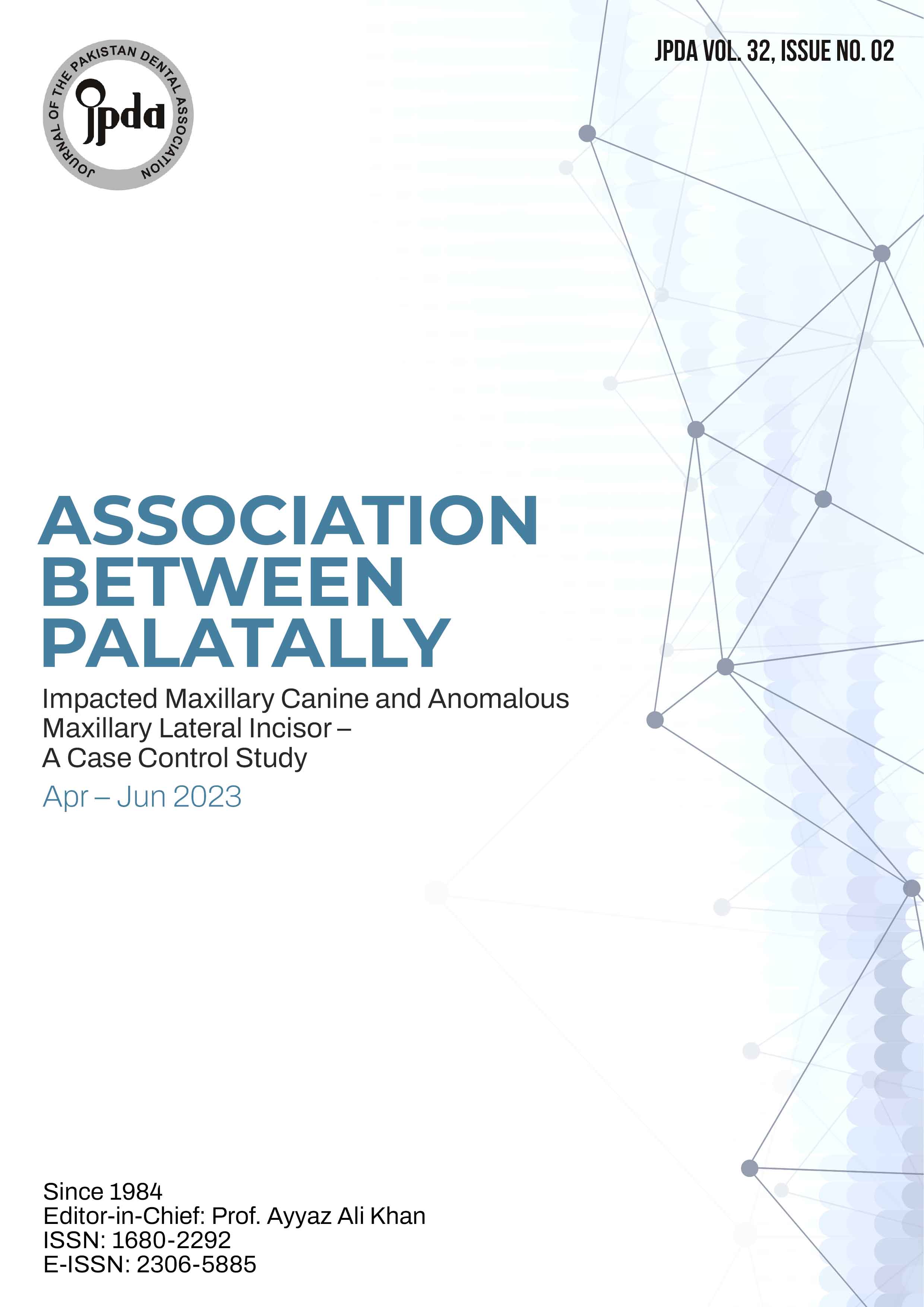
The inclusion criteria were participants without syndromes, palatal canine impaction (cases only), and age range from 12 to 30 year. Subjects with buccal impacted canine, history of previous orthodontic treatment and non-Pakistani nationals were excluded. Participant’s OPG and periapical X-rays were used to diagnose PIC by horizontal parallax technique. Dental anomalies in upper lateral incisor like missing and peg shaped were further diagnosed by using casts and OPGs
The data were analyzed using SPSS 22. Mean and SD were calculated for continuous data like age and frequency and percentages for categorical data like gender and anomalies of upper lateral incisors. Chi-square /Fisher exact test was run to compare anomalies of upper lateral incisors between cases and control. To quantify the degree of association the odds ratio with 95% confident intervals was calculated by binary logistic regression between dependent variable (anomalies in lateral incisor) and independent variable (palatally impacted canine). P<0.05 was considered as significant level.
RESULTS:
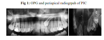
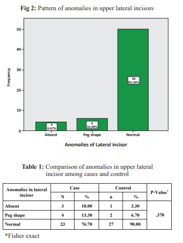
The mean age of the study participants was 18.916±4.3 years and ranging from 13 to 29 years. Females (n=33, 55%) were more than males (n=27, 45%). The frequency of missing lateral incisor was higher in cases (n=3, 10%) than control (n=1, 3.3%). Similarly peg shape laterals were more in cases
(n=4, 13.3%) than controls (n=2, 6.7%). However the association was not statistically significant (P=0.37).(Fig 2)
DISCUSSION:
This case control study was aimed to determine the association between palatally impacted maxillary canine and anomalies in upper lateral incisor. Our findings showed that though the frequency of lateral incisor’s anomalies are higher in participants with palatally impacted canine than normal but this association was not statistically significant (P=0.37). The logistic regression analysis showed that odds of having lateral incisors anomalies were about three times higher but this was not significant statistically. Our results showed that association between anomalies in lateral incisor and palatally impacted canine was positive (OR=2.73) and not statistically significant. According to guidance theory maxillary canine erupt by taking guidance from root of lateral incisor.11 A study conducted by Laganà et al.12 in Italy on 336 subjects on association of displaced maxillary canine and anomalous lateral incisors. They found a statistically significant and positive association (OR=1.139, 95%= 1.43-4.15%). The non-significance of our results can be due to less number of participants in our study. Other studies also found evidence for association of PIC with upper laterals agenesis.13,14
In our study missing lateral incisor was 10% in controls and 13.3% in cases. A previous study conducted in Rawalpindi reported that agenesis of lateral incisor was 6.7% in controls and 13.3% in cases. These results support our findings.15 Another study by Garib et al.16 reported that there was 18.9% upper lateral incisor hypodontia in cases with unilateral palatal canine impactions. Another investigation by Zilberman et al.17 reported 5.5% congenitally absent upper laterals in cases with PIC. Peck and Peck found 3.4% missing maxillary lateral incisors in association with palatally impacted canines.18 Another study found 7.23% congenitally absent upper laterals incisors with palatally impacted canines.19
The current study showed that peg shaped laterals were more in cases (13.3%) than controls (6.7%). However the association was not statistically significant (P=0.37). Similar results were reported by previous study conducted in Pakistan.15
CONCLUSION:
Within the limits of this study we can conclude that though the frequency of anomalous maxillary lateral incisors is higher in participants with palatal impacted canine than controls but this association was not statistically significant.
CONFLICT OF INTEREST:
None declared
REFERENCES:
1. Ngo CTT, Fishman LS, Rossouw PE, Wang H, Said O. Correlation between panoramic radiography and cone-beam computed tomography in assessing maxillary impacted canines. Angle Orthod. 2018;88:384- 9.
https://doi.org/10.2319/103117-739.1
2. Yan B, Sun Z, Fields H, Wang L. Maxillary canine impaction increases root resorption risk of adjacent teeth: a problem of physical proximity. Am J Orthod Dentofacial Orthop. 2012;142:750-7.
https://doi.org/10.1016/j.ajodo.2012.07.016
3. Dogramaci EJ, Sherriff M, Rossi-Fedele G, McDonald F. Location and severity of root resorption related to impacted maxillary canines: a cone beam computed tomography (CBCT) evaluation. Austral Orthod J. 2015;31:49-59.
https://doi.org/10.21307/aoj-2020-140
4. Cabay RJ. An overview of molecular and genetic alterations in selected benign odontogenic disorders. Archive Pathol Labor Med. 2014;138:754-8.
https://doi.org/10.5858/arpa.2013-0057-SA
5. Agrawal P, Manohar S, Thorne-Lyman AL, Angela K, Shrestha B, Klemm RD, et al. Prevalence of damaged and missing teeth among women in the southern plains of Nepal: Findings of a simplified assessment tool. PloS one. 2019;14:e0225192.
https://doi.org/10.1371/journal.pone.0225192
6. Cruz RM. Orthodontic traction of impacted canines: Concepts and clinical application. Dent Press J Orthod. 2019;24:74-87.
https://doi.org/10.1590/2177-6709.24.1.074-087.bbo
7. Alqahtani H. Management of maxillary impacted canines: A prospective study of orthodontists’ preferences. Saudi Pharmac J. 2021;29:384-90.
https://doi.org/10.1016/j.jsps.2021.03.010
8. Manne R, Gandikota C, Juvvadi SR, Rama HRM, Anche S. Impacted canines: Etiology, diagnosis, and orthodontic management. J Pharm
Bioallied Sci. 2012;4(Suppl 2):S234.
https://doi.org/10.4103/0975-7406.100216
9. Jena AK, Duggal R. The pattern of maxillary canine impaction in relation to anomalous lateral incisors. J Clin Pediatr Dent. 2010;35:37- 40.
https://doi.org/10.17796/jcpd.35.1.uh4vm67264vv4762
10. Becker A, Smith P, Behar R. The incidence of anomalous maxillary lateral incisors in relation to palatally-displaced cuspids. Angle Orthod. 1981;51:24-9.
11. Bertl MH, Foltin A, Lettner S, Giannis K, Gahleitner A, Bantleon H-P, et al. Association between maxillary lateral incisors’ root volume and palatally displaced canines: An instrumental variables approach to the guidance theory. Angle Orthod. 2018;88:719-25.
https://doi.org/10.2319/020818-107.1
12. Laganà G, Venza N, Lione R, Chiaramonte C, Danesi C, Cozza P. Associations between tooth agenesis and displaced maxillary canines: a cross-sectional radiographic study. Prog Orthod. 2018;19:1- 6.
https://doi.org/10.1186/s40510-018-0226-0
13. Krishnaveni S, Reddy YM, Sreekanth C, Reddy BV, Kranthi G, Raj P, et al. Nasal Integument as an Indicator of Maxillary Skeletal
Pattern. Int J Oral Health Med Res 2017;3:31-5.
14. Amit G, Pankaj B, Suchinder S, Parul B. Periodontally accelerated osteogenic orthodontics (PAOO)-a review. 2012.
https://doi.org/10.4317/jced.50822
15. Enlow DH, Hans MG. Essentials of facial growth Philadelphia: WB Saunders; 2008.
16. Scott J. The cranial base. Am J Phys Anthropol. 1958;16:319-48.
https://doi.org/10.1002/ajpa.1330160305
17. Bhushan R, Kumar S, Chauhan AK, Mohan S, Shekhar M, Narnoly A. Assessment of the relationship between maxillary rotation and nasal morphology in males. Contemp Clin Dent. 2015;6(Suppl 1):S12-S7.
https://doi.org/10.4103/0976-237X.152931
18. Peck S, Peck L, Kataja M. Prevalence of tooth agenesis and pegshaped maxillary lateral incisor associated with palatally displaced canine (PDC) anomaly. Am J Orthod Dentofac Orthop 1996;110: 441-43.
https://doi.org/10.1016/S0889-5406(96)70048-3
19. Leifert S, Jonas IE. Dental anomalies as a microsymptom of palatal canine displacement. J Orofac Orthop 2003;64:108-20.
https://doi.org/10.1007/s00056-003-0222-x