
Lack of Association of Palataly Impacted Canines with Maxillary Arch Width and Lateral Incisor Anomalies
INTRODUCTION
The most frequently impacted tooth in the dental arch after the third molars is the maxillary canine. The incidence of its impaction is between 1 and 2.5 %. Impactions of canine can be either buccal or lingual.1 Around 85% of impactions are palatal while the remaining 15% are buccal. Canine impaction occurs twice as more in the maxilla than in the mandible. In subjects with impacted maxillary canines, 8% of impactions occur bilaterally.2
The etiology of palatal canines is accepted to be multifactorial and of a complex nature. The two accepted theories for this anomaly are guidance theory and genetic theory. The guidance theory stresses on the impact of lateral incisors on the eruption of palatal canines and also states that absent, peg or anomalous lateral incisors lead to the impaction of maxillary canines. The genetic theory explains the occurrence of different dental anomalies that occur along with palataly impacted canines e.g. infraoccluded deciduous molars and ectopic eruption of maxillary 1st molars.3,4
Maxillary width is usually decreased in patients with buccaly impacted canines5 but the relationship between width of maxilla and palatal canines is questionable. McConell in a study implicated maxillary width deficiency as a factor for palatal canine displacement.6 On the other hand, Langberg and Peck found no statistical significant difference in the anterior and posterior maxillary arch widths in patients with palataly displaced canines.1,2,7
There is compelling evidence that palataly displaced canines are associated with agenesis of lateral incisors as well as peg shaped lateral incisors.8 Becker and Smith in their study indicated that the absence of lateral incisor could lead to palatal canine impaction.9
Al Nimri in his study found smaller mesiodistal width and short roots of lateral incisors in association with palatal canines.10 Contrary to the aforementioned evidence, there are studies that reflect a weak association between palataly impacted canines and lateral incsiors. Mossey et al in their study showed a weak support between impacted canines and absence of adjacent lateral incisors.11 Jena and Duggal in their study also found no positive association between lateral incisor anomalies and canine impaction.12
As such an association has not been checked on Pakistani population, our aim with this study was to explore a relationship between palatally impacted canines and maxillary interarch widths at the level of first premolar and first molars, and also their possible relation with anomalies of lateral incisors (agenesis/ peg laterals).
METHODOLOGY
This cross sectional study was conducted on the orthopantomograms and dental casts of the patients coming for orthodontic treatment at the Dental OPD of Bahria University Medical and Dental College between the periods of 2016 to 2019. An institutional ethical committee approved the protocol of study (letter no ERC 39/2019). The sample was selected via convenience sampling. The sample size was calculated with the OPENEPI online software. With a standard of 95% confidence interval and 5% error size, the proportion of subjects and controls was found to be 60 (30 subjects and 30 controls). The experimental group comprised subjects diagnosed with palatal canine impaction while the control group included subjects without palatal impactions.
The inclusion criteria for experimental subjects included:
a) Nonsyndromic subjects
b) Unilateral palatal canine impaction
c) Age group: 12 to 30 years
The exclusion criteria for experimental subjects are:
a) Missing 1st premolars
b) Missing 1st molars
c) Severely mesially rotated 1st molars
d) Previous orthodontic treatment
The patients were documented with OPG’s and Periapical

FIG 1
x rays (horizontal parallax technique) for the presence of palatal canine. Pretreatment dental casts of the patients were used to assess maxillary first inter-premolar and first intermolar widths. These readings would be used to determine the maxillary transverse width. The data was collected by a single examiner who was calibrated to make these measurements by placing the caliper tips into the deepest portion of the central fossae of the upper first premolars at their junctions with the most lingual aspect of the buccal
cusp. The same examiner also recorded the intermolar width by placing the caliper tips into the deepest portion of the central fossae at its junction with the most lingual aspect of
the mesio-buccal cusp. All dental cast measurements were made using a digital boley gauge.
The dental anomalies to be included in the experimental and control group included lateral incisor’s agenesis /peg laterals.
STATISTICAL ANALYSIS
Data analysis was done by using SPSS version 23. Fisher exact test was used to determine the co-relation between impacted canines and missing or anomalous lateral incisors.
To observe the correlation of impaction with intermolar and interpremolar width (mm), independent sample t test was applied. P value <0.05 was considered as statistically
significant.
RESULTS
A total of 60 patients (30 experimental and 30 controls) were examined in this study. Mean age in experimental subjects was 17.93±5.23 years, whereas in controls it was found to be 18.30±3.90 years. The presence/ absence of lateral incisors as well as anomalies are presented in Table 1. There were 25 (83.3%) experimental subjects who had normal shaped lateral incisors, whereas in controls normal lateral incisors were present in
Table 1: Intergroup comparison of the number of lateral incisor anomalies

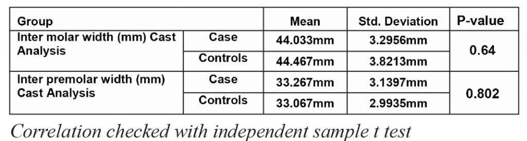
Table 2: Intergroup comparisons of maxillary intermolar and interpremolar widths (in mm)
28 (93.3%) cases. Peg lateral incisor, an anomaly of lateral incisors was found in only 1 (3.3%) experimental subject whereas none in controls with non-significant p-value of 0.339.
Mean Inter molar width (mm) in experimental subjects was found to be 44.03±3.29 whereas in controls it was found to be 44.46±3.82 with non-significance p-value of
0.64. Mean Inter premolar width (mm) in experimental cases was found to be 33.26±3.13 whereas in controls 33.06±2.99 was observed with non-significant p-value of 0.802. (Table 2)
DISCUSSION
Impaction of the maxillary canine is a common occurrence and is encountered frequently by orthodontists as well as general dentists. It has a prevalence of 1-2.5% and is the second only to developmental disturbance in third molars.1,13 Diagnosis of this condition at an early age along with preventive measures such as rapid maxillary expansion, extraction of deciduous canine and first molars, transpalatal arches as well as cervical headgear can aid in spontaneous eruption of these teeth. If the palatal canine is left untreated, it can result in various side effects of which the noteworthy ones include cyst formation, root resorption of adjacent teeth and development of malocclusion.9
In our study, lateral incisor hypodontia was found in only 13% out of 30 experimental cases with palatal canine impactions while in controls; only 6.7% of laterals were absent. Only one case of peg lateral incisor (3.3%) was found in experimental subjects while none was reported in controls. Even though some studies have established a correlation between impacted teeth with dental anomalies, our study showed no correlation. Our study was in accordance with the study of Garib et al who had maxillary lateral incisor agenesis (18.9%) for subjects with unilateral palatal canine impactions.14 Zilberman et al in their study showed similar results with only 5.5% lateral incisors missing in patients with palatal canines whereas the percentage of peg laterals was 13%.15 Peck and Peck in their study also found lateral incisor hypodontia to be 3.4% in cases with palatal canines.16 Leifert and Jonas found similar results to our study with 7.23% hypodontia of lateral incisors.11 From the results of our study, we found that the maxillary transverse dimension measured as first interpremolar and intermolar width was similar in experimental cases as well as in controls. We found the mean intermolar width to be 44 mm in both subjects and controls with a p value of 0.64 whilst the mean interpremolar width was 33 mm in both groups with a p value of 0.8. Our study was in accordance with the study of Gull and Mushtaq who also found the intermolar width to be 44mm and the interpremolar width to be 35 mm with no significant correlation to the impacted teeth.17 Naoumova and Alfaro in their study also found no difference between arch width at the molars in both impaction cases and controls.18 Ariza et al in their study found contrasting results and showed that the transverse dimensions of the first molar and first premolar were smaller in subjects with impacted maxillary canines when compared with subjects without impaction.19
From our study and the results of various other researches, we can confirm that dental anomalies like agenesis of lateral incisors, peg lateral incisors as well as maxillary skeletal width do not correlate positively with the presence of palatal canine impactions. Further investigation into the etiology of palataly impacted canines is needed before clinical recommendations can be made to find more factors that correlate positively with palatal canines.
CONCLUSION
Our study did not show any relationship between palataly impacted canines with anomalous and missing incisors or maxillary width.
CONFLICT OF INTEREST
None declared
REFERENCES
- K. Al-Nimri and T. Gharaibeh. Space conditions and dental and occlusal features in patients with palataly impacted maxillary canines: an etiological study. Europ J Orthod 27;2005:461-65. https://doi.org/10.1093/ejo/cji022
- Manne R, Gandikota C, Juvvadi SR, Medapati Rama H, Anche S.Impacted canines: Etiology, diagnosis, and orthodontic management.J Pharm Bioall Sci 2012;4:234-38. https://doi.org/10.4103/0975-7406.100216
- Saiar M, Rebellato J, and Sheats RD. Palatal displacement of canines and maxillary skeletal width. Am J Orthod Dentofacial Orthop 2006;129:511-19 https://doi.org/10.1016/j.ajodo.2005.03.021
- Hong W, Radfar R, Chung C. Relationship between the maxillary transverse dimension and palataly displaced canines: A cone beam computed tomographic study. Angle Orthod. 2015:85;440-445. https://doi.org/10.2319/032614-226.1
- Ariza NA, Schilling J, Guillen LEA, Mora GAR, Cardenas YAR, and Castillo AAD. Maxillary transverse dimensions in subjects with and without impacted canines: A comparative cone-beam computed tomography study. Am J Orthod Dentofacial Orthop 2018;154:495- 503 https://doi.org/10.1016/j.ajodo.2017.12.017
- McConell TL, Hoffman DL, Forbes DP, Janzen EK, Weintraub NH. Maxillary canine impaction in patients with transverse maxillary` deficiency. ASDC J Dent Child. 1996;63:190-95.
- Fattahi H, Ghaeed F, Alipour A. Association betweenmaxillary canine impaction and arch dimensions. Aust Orthod J. 2012;28:57-62
- Mercuria E; CassettM;Cavallini C; Vicari D; Leonardi R; Barbato E. Dental anomalies and clinical features in patients with maxillary canine impaction. A retrospective study. Angle Orthod. 2013;83:22- 28. https://doi.org/10.2319/021712-149.1
- Becker A, Smith P, Behar R. The incidence of anomalous lateral incisors in relation to palataly displaced cuspids. Angle Othod. 1981;51:24-9.
- Al-Nimri KS, Bsoul E. Maxillary palatal canine impaction displacement in subjects with congenitally missing maxillary lateral incisors. Am J OrthodDentofacialOrthop. 2011;140:81-86. https://doi.org/10.1016/j.ajodo.2009.11.016
- Mossey PA, Campbell HM, Luffingham JK. The palatal canine and the adjacent lateral incisor: a study of a west of Scotland population. Br J Orthod. 1994:21:169-74. https://doi.org/10.1179/bjo.21.2.169
- Jena AK, Duggal R. The pattern of maxillary canine impaction in relation to anomalous lateral inciosrs. 13 13 Leifert S, Jonas IE. Dental Anomalies as a Microsymptom of Palatal Canine Displacement. J Orofac Orthop 2003;64:108-20. https://doi.org/10.1007/s00056-003-0222-x
- Leifert S, Jonas IE. Dental Anomalies as a Microsymptom of Palatal Canine Displacement. J Orofac Orthop 2003;64:108-20. https://doi.org/10.1007/s00056-003-0222-x
- Garib DG, Alencar BM, Lauris JRP, Bacetti T. Agenesis of maxillary lateral incisors and associated dental anomalies. Am J Orthod Dentofacial Orthop2010;137:732.e1-732. https://doi.org/10.1016/j.ajodo.2009.12.024
- Zilberman Y, Cohen B, Becker A. Familial trends in palatal canines, anomalous lateral incisors, and related phenomena. Europ J Orthod 1990;12:135-39. https://doi.org/10.1093/ejo/12.2.135
- Peck S, Peck L, Kataja M. Prevalence of tooth agenesis and pegshaped maxillary lateral incisor associated with palatally displaced canine (PDC) anomaly. Am J Orthod Dentofac Orthop 1996;110:441- 43. https://doi.org/10.1016/S0889-5406(96)70048-3
- Gull MAB, Mushtaq M, Maqbool S. Palatal Displacement of Maxillary Canines and Maxillary Transverse Dimensions. Annals Int Med Dent Res. 2018;4:10. https://doi.org/10.21276/aimdr.2018.4.2.DE3
- Naoumova J, AlfaroGE; Peck S. Space conditions, palatal vault height, and tooth size in patients with and without palatally displaced canines:Angle Orthod 2018;88:726-32. https://doi.org/10.2319/120717-843.1
- Ariza NA, Schilling J, Guillen LEA, Mora GAR, Cardenas YAR, Castillo AAD. Maxillary transverse dimensions in subjects with and without impacted canines: A comparative cone-beam computed tomography study. Am J Orthod Dentofacial Orthop 2018;154:495-50 https://doi.org/10.1016/j.ajodo.2017.12.017